Leg Bones Diagram / Appendicular Skeleton Learn Skeleton Anatomy - Lower jaw (mandible) collar bone.. Visit kenhub for more skeletal system quizzes. The bones involved in it, however, are only the femur and the tibia, although the smaller bone of the leg, the fibula, is carried along in the movements of flexion, extension, and slight rotation that this joint. Quizzes on human skeletal system anatomy, bone anatomy, and bone markings. The knee joint is the largest joint in the body and is primarily a hinge joint although some sliding and rotation. These simple labelled diagrams of the bones of the lower legs and feet and the bones of the arms and hands this diagram shows the skeletal structure of the leg (anterior view) and foot (dorsal view).
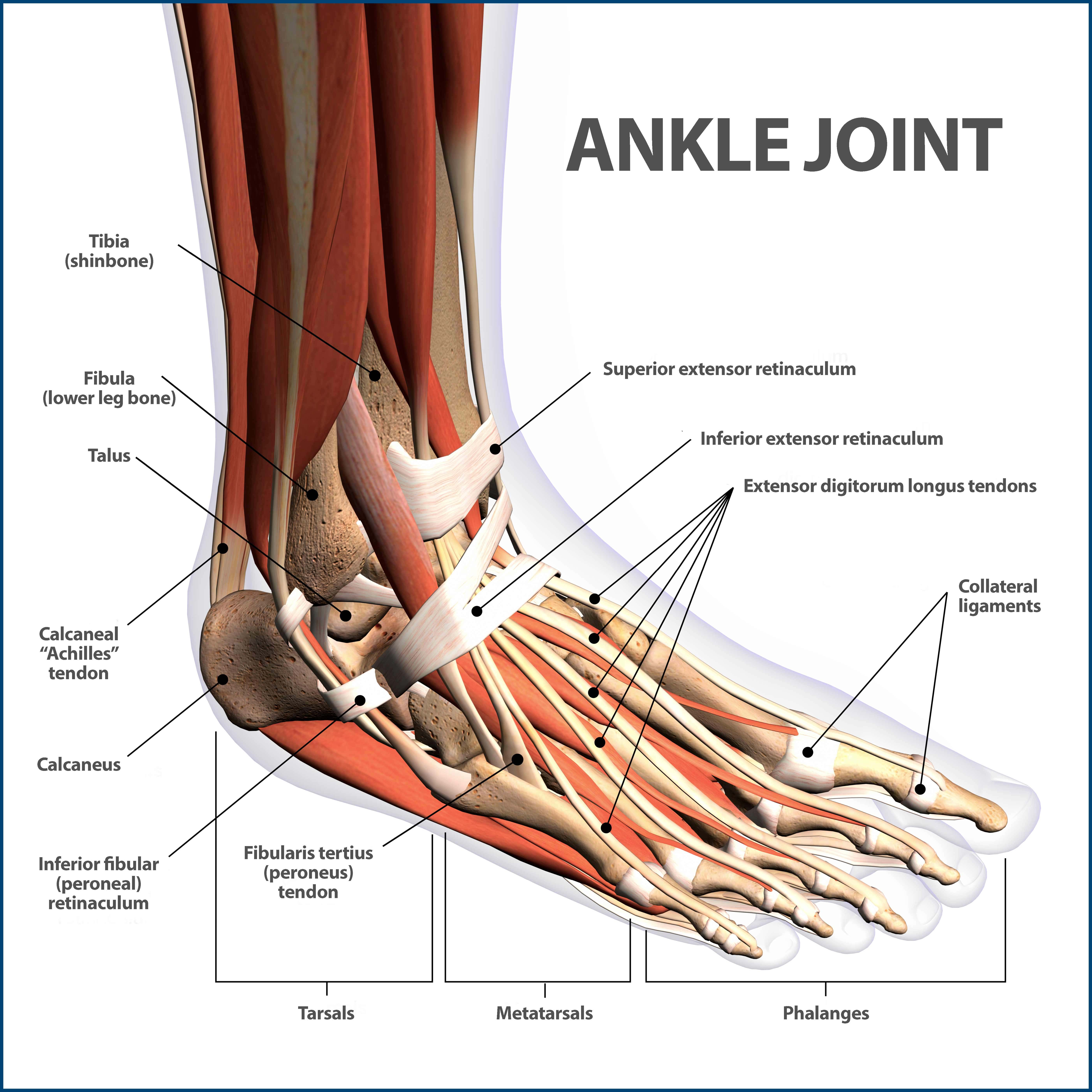
When you stand or walk, all the weight of your upper body rests on them. Click now to learn more about the bones, muscles, and soft tissues tibia: Your leg bones are the longest and strongest bones in your body. Your legs are two of your most important body parts. The foot bones shown in this diagram are the talus, navicular, cuneiform, cuboid, metatarsals.
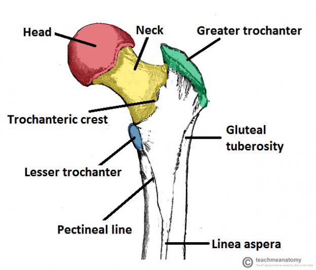
Ankle Fractures Broken Ankle Florida Orthopaedic Institute from www.floridaortho.com Each leg is made up of four bones. Learn how to draw the femur, patella, tibia, and fibula in this lesson! This bright worksheet helps your child bring these technical terms down to size. Most bones (particularly the long bones of the arms and legs — which make up the appendicular skeleton) have a hard outer shell known as cortical bone. The bone that goes from your pelvis to your knee is called the femur (say: High resolution textures and displacement included. Bone surfaces at synovial joints are protected by a coating of articular cartilage. At the same time, the bones and joints of the leg and foot must be strong enough to support the body's weight while remaining flexible enough for movement and balance.
The musculoskeletal segment of the leg, including the foot bones (ankle, heel bone, toe bones), fibula and tibia, knee, femur and femoral neck, hip and sacrum as well as the third, fourth.
It expands at the proximal and distal ends, articulating at the knee and ankle joints respectively. Each leg is made up of four bones. He'll boost his body knowledge as he matches up the names of the bones with their proper places on the leg diagram. Learn vocabulary, terms and more with flashcards, games and other study tools. Most bones (particularly the long bones of the arms and legs — which make up the appendicular skeleton) have a hard outer shell known as cortical bone. Health diagram bone skeleton leg knee science anchor chart human human body. Quizzes on human skeletal system anatomy, bone anatomy, and bone markings. The foot bones shown in this diagram are the talus, navicular, cuneiform, cuboid, metatarsals. Your leg bones are the longest and strongest bones in your body. At the same time, the bones and joints of the leg and foot must be strong enough to support the body's weight while remaining flexible enough for movement and balance. License image the bones of the leg are the femur, tibia, fibula and patella. The bones and joints in the feet experience wear and tear, so conditions that cause damage to the it is usually the result of a muscle imbalance when the long muscles of the lower leg overpower the. This long bone connects with the knee at one end and the next to the tibia is the fibula, the thinner, weaker bone of the lower leg.
Cheek bone (zygoma) upper jaw (maxilla). They allow you to move and provide support for your upper body. The knee joint is the largest joint in the body and is primarily a hinge joint although some sliding and rotation. It expands at the proximal and distal ends, articulating at the knee and ankle joints respectively. The bones and joints in the feet experience wear and tear, so conditions that cause damage to the it is usually the result of a muscle imbalance when the long muscles of the lower leg overpower the.
Diagram Upper Leg Bone Diagram Labeled Full Version Hd Quality Diagram Labeled Alphawiring Mondemodexl Fr from jb004.k12.sd.us Skeleton leg ankle joints and toe phalanges, cuboid, metatarsal, navicular and cuneiform bones, hand drawn dorsal view of foot. The knee joint is the largest joint in the body and is primarily a hinge joint although some sliding and rotation. High resolution textures and displacement included. The human leg, in the general word sense, is the entire lower limb of the human body, including the foot, thigh and even the hip or gluteal region. Learn how to draw the femur, patella, tibia, and fibula in this lesson! Visit kenhub for more skeletal system quizzes. Diagram and names of leg bones, diagram of foot and leg bones, diagram of leg bones, diagram of lower leg related posts of diagram of leg bones. Bones of the leg and foot, lower leg bone anatomy, leg bones anatomy, leg muscles, leg bones diagram, leg bone structure, leg anatomy muscles, parts of the lower leg.
The bones of the leg are the femur, tibia, fibula and patella.
Synovial joints are often supported and reinforced by surrounding ligaments, which limit movement to prevent injury. You'll learn about the muscles, bones, and other structures of each area of the leg. Click now to learn more about the bones, muscles, and soft tissues tibia: The foot bones shown in this diagram are the talus, navicular, cuneiform, cuboid, metatarsals and calcaneus. The bones of the leg are the femur, tibia, fibula and patella. Diagram and names of leg bones, diagram of foot and leg bones, diagram of leg bones, diagram of lower leg related posts of diagram of leg bones. Quizzes on human skeletal system anatomy, bone anatomy, and bone markings. Health diagram bone skeleton leg knee science anchor chart human human body. At the same time, the bones and joints of the leg and foot must be strong enough to support the body's weight while remaining flexible enough for movement and balance. The human leg, in the general word sense, is the entire lower limb of the human body, including the foot, thigh and even the hip or gluteal region. Time to jump right into the biggest and strongest bones in the human body. Bones of the leg and foot, lower leg bone anatomy, leg bones anatomy, leg muscles, leg bones diagram, leg bone structure, leg anatomy muscles, parts of the lower leg. The knee joint is the largest joint in the body and is primarily a hinge joint although some sliding and rotation.
Master leg and knee anatomy using our topic page. The bones and joints in the feet experience wear and tear, so conditions that cause damage to the it is usually the result of a muscle imbalance when the long muscles of the lower leg overpower the. Your leg bones are the longest and strongest bones in your body. This long bone connects with the knee at one end and the next to the tibia is the fibula, the thinner, weaker bone of the lower leg. Your legs are two of your most important body parts.
Bones Of The Lower Limb Teachmeanatomy from teachmeanatomy.info Synovial joints are often supported and reinforced by surrounding ligaments, which limit movement to prevent injury. The largest and most medial leg bone, forming both the knee and ankle joints. Cheek bone (zygoma) upper jaw (maxilla). High quality realistic skeleton legs. Click now to learn more about the bones, muscles, and soft tissues tibia: They allow you to move and provide support for your upper body. It expands at the proximal and distal ends, articulating at the knee and ankle joints respectively. He'll boost his body knowledge as he matches up the names of the bones with their proper places on the leg diagram.
The largest and most medial leg bone, forming both the knee and ankle joints.
Your leg bones are the longest and strongest bones in your body. Most bones (particularly the long bones of the arms and legs — which make up the appendicular skeleton) have a hard outer shell known as cortical bone. This bright worksheet helps your child bring these technical terms down to size. Synovial joints are often supported and reinforced by surrounding ligaments, which limit movement to prevent injury. The bones involved in it, however, are only the femur and the tibia, although the smaller bone of the leg, the fibula, is carried along in the movements of flexion, extension, and slight rotation that this joint. Click now to learn more about the bones, muscles, and soft tissues tibia: The second largest bone in body is the tibia, also called the shinbone. Master leg and knee anatomy using our topic page. The human leg, in the general word sense, is the entire lower limb of the human body, including the foot, thigh and even the hip or gluteal region. The foot bones shown in this diagram are the talus, navicular, cuneiform, cuboid, metatarsals and calcaneus. Visit kenhub for more skeletal system quizzes. Diagram and names of leg bones, diagram of foot and leg bones, diagram of leg bones, diagram of lower leg related posts of diagram of leg bones. The foot bones shown in this diagram are the talus, navicular, cuneiform, cuboid, metatarsals.

Komentar
Posting Komentar